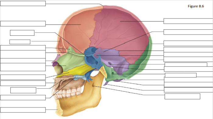
Prepare to delve into the depths of the midsagittal view of the skull, a captivating journey that unravels the intricate tapestry of our protective cranial dome. This vantage point offers a unique perspective, akin to a window into the skull’s hidden architecture, revealing its remarkable landmarks, sutures, sinuses, and more.
Our exploration begins with a comprehensive examination of the anatomical landmarks that grace this view, each serving a vital role in the skull’s overall structure and function. We will then venture into the fascinating realm of sutures and fontanelles, unraveling their significance in the skull’s development and adaptation throughout life.
Anatomical Landmarks of the Midsagittal View
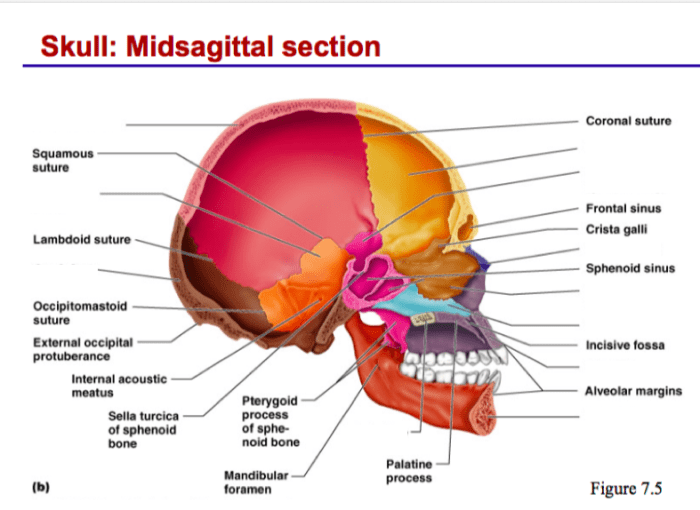
The midsagittal view of the skull is an anatomical orientation that divides the skull into left and right halves. It is a useful view for examining the overall structure of the skull and the relationships between its various components.
The major anatomical landmarks visible in the midsagittal view include:
- Frontal bone: Forms the forehead and the upper part of the eye sockets.
- Parietal bone: Forms the upper and lateral sides of the skull.
- Occipital bone: Forms the back of the skull.
- Sphenoid bone: Forms the base of the skull and supports the pituitary gland.
- Ethmoid bone: Forms the roof of the nasal cavity and the medial walls of the eye sockets.
- Nasal bone: Forms the bridge of the nose.
- Maxilla: Forms the upper jaw.
- Mandible: Forms the lower jaw.
- Teeth: Embedded in the maxilla and mandible.
- Tongue: Occupies the floor of the oral cavity.
These landmarks provide a framework for understanding the overall structure of the skull and its relationship to the other structures of the head and neck.
Sutures and Fontanelles
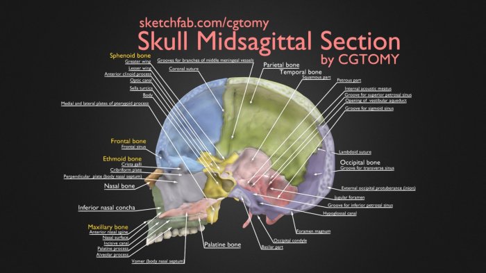
Sutures are immovable joints that connect the bones of the skull. Fontanelles are unossified areas between the bones of the skull that are filled with fibrous connective tissue. Both sutures and fontanelles play a vital role in the development and growth of the skull.
During early life, the skull is highly flexible and adaptable. This is due in part to the presence of sutures and fontanelles. The sutures allow the bones of the skull to move slightly, which helps to accommodate the growth of the brain.
The midsagittal view of the skull, with its distinct profile, provides a wealth of information about the individual’s facial structure. Just as the examen de señales de transito helps drivers navigate the complexities of the road, the midsagittal view guides anthropologists in understanding the evolutionary and functional aspects of the human skull.
The fontanelles provide even greater flexibility, as they allow the bones of the skull to overlap. This overlapping helps to protect the brain from injury during birth and during the early years of life.
Major Sutures and Fontanelles Visible in the Midsagittal View
- Sagittal suture:This suture runs along the midline of the skull, connecting the two parietal bones.
- Coronal suture:This suture runs across the top of the skull, connecting the frontal bone to the two parietal bones.
- Lambdoid suture:This suture runs along the back of the skull, connecting the occipital bone to the two parietal bones.
- Anterior fontanelle:This fontanelle is located at the junction of the sagittal and coronal sutures. It is the largest fontanelle and closes by about 18 months of age.
- Posterior fontanelle:This fontanelle is located at the junction of the sagittal and lambdoid sutures. It is smaller than the anterior fontanelle and closes by about 6 months of age.
As the child grows, the sutures and fontanelles gradually close. This process is complete by about 2 years of age. The closure of the sutures and fontanelles helps to stabilize the skull and protect the brain from injury.
Cranial Vault and Base: Midsagittal View Of The Skull
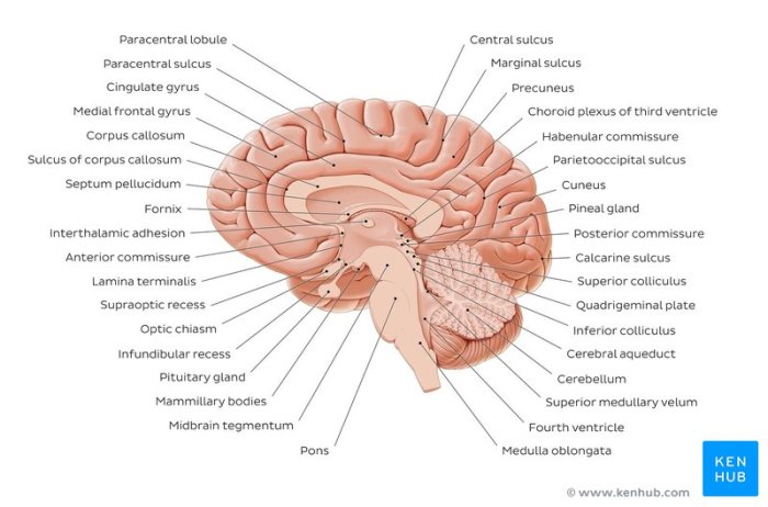
The cranial vault is the dome-shaped upper portion of the skull that encloses and protects the brain. It consists of eight bones: the frontal bone, two parietal bones, two temporal bones, the occipital bone, and the sphenoid bone. These bones are joined together by immovable joints called sutures.The
cranial vault provides protection for the brain from mechanical injury, such as blows to the head. It also helps to regulate the temperature of the brain and provides attachment points for muscles that move the head and face.The cranial base is the lower portion of the skull that supports the brain and provides passage for nerves and blood vessels.
It consists of the occipital bone, the sphenoid bone, the temporal bones, and the ethmoid bone. The cranial base is divided into three regions: the anterior cranial fossa, the middle cranial fossa, and the posterior cranial fossa.The anterior cranial fossa contains the frontal lobes of the brain.
The middle cranial fossa contains the temporal lobes of the brain. The posterior cranial fossa contains the cerebellum and the brainstem.
Paranasal Sinuses
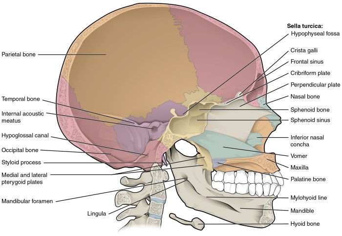
The midsagittal view of the skull reveals several paranasal sinuses, which are air-filled cavities within the facial bones. These sinuses include the frontal sinuses, ethmoid sinuses, maxillary sinuses, and sphenoid sinuses.
Paranasal sinuses play a crucial role in respiration and drainage. They lighten the skull, warm and moisten inhaled air, and contribute to the production of mucus that traps dust and other particles. The sinuses also provide resonance for the voice.
Clinical Significance
Paranasal sinus infections, also known as sinusitis, are common conditions that can cause pain, swelling, and difficulty breathing. Sinusitis can be caused by viruses, bacteria, or allergies. Treatment typically involves antibiotics, decongestants, and pain relievers.
Clinical Applications
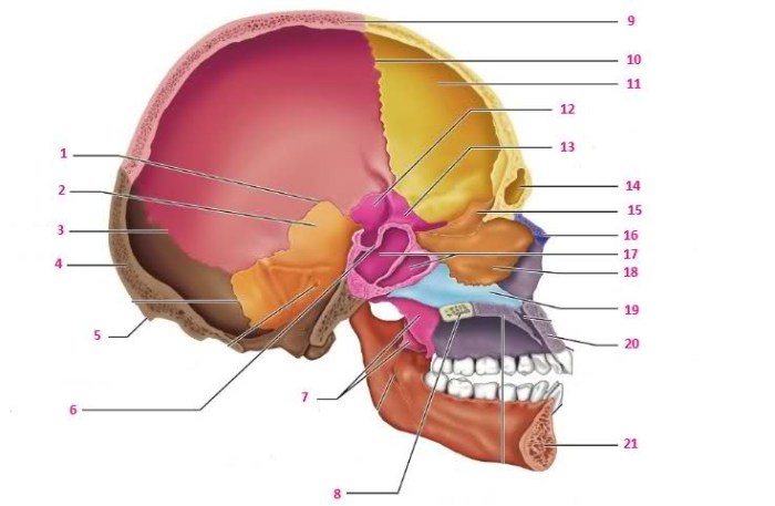
The midsagittal view provides valuable information for diagnosing and managing various skull-related conditions.
Diagnosing and Managing Skull Fractures, Midsagittal view of the skull
The midsagittal view allows for the assessment of skull fractures, including their location, extent, and displacement. By visualizing the alignment of the cranial bones, it helps determine the severity of the injury and guide treatment decisions.
Assessing Intracranial Pressure and Detecting Brain Abnormalities
The midsagittal view can provide indirect evidence of increased intracranial pressure by showing widening of the sutures, particularly the sagittal suture. Additionally, it can detect brain abnormalities such as tumors, cysts, or herniations, which may appear as shifts in the midline structures.
Surgical Planning and Monitoring
The midsagittal view is crucial in surgical planning, especially for procedures involving the skull base. It provides a clear visualization of the anatomical relationships between the skull bones, dura mater, and neurovascular structures, aiding in the selection of the optimal surgical approach and minimizing potential complications.
Essential Questionnaire
What is the midsagittal view of the skull?
The midsagittal view is a median section of the skull that divides it into left and right halves, providing a comprehensive view of its internal structures.
What are the major anatomical landmarks visible in this view?
The midsagittal view reveals key landmarks such as the frontal bone, parietal bone, occipital bone, sphenoid bone, ethmoid bone, nasal bone, and mandible.
What is the clinical significance of the midsagittal view?
The midsagittal view is crucial for diagnosing and managing skull fractures, assessing intracranial pressure, detecting brain abnormalities, and guiding surgical planning.
